The Vestibular System

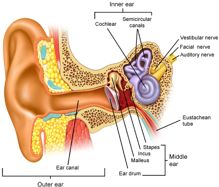
Before going into the details of various vestibular disorders, it is important to understanding the anatomical components involved in the human inner ear. The inner ear consists of a bony labyrinth, which is a hollow cavity in the temporal bone of the skull, with a system of passages that makes up two main functional divisions. The first division is the cochlea (responsible for hearing), where sound pressure patterns are converted from the outer ear into electrochemical impulses which are passed on to the brain via the auditory nerve. The second division, the vestibular system (responsible for balance), detects linear acceleration and head orientation relative to gravity (Dohlman, 1944). The vestibular system consists of the otolith organs (utricle and saccule), as well as three semicircular canals (anterior, posterior and horizontal). The semicircular canals are a network of tubes, oriented at almost right angles to each other and filled with endolymph fluid (Dohlman, 1944). A swelling exists on one end of each semicircular canal called the ampulla. Inside the ampulla lies a gelatinous mass called the cupula and hair cells (Dohlman, 1944). As the position of the head changes, the endolymph flow within the inner ear moves, the cupula is deflected, and tiny sensory hair cells are stimulated, signaling head movement. (Dohlman, 1944).
Information is passed through the vestibular nerve to the brain about head rotation movements, linear movements as well as static postures of the head relative to gravity. In response, the brain sends commands to your eyes for improved vision and to your skeletal muscles for greater sitting balance, standing balance and movement stability.
Within the otolith organs lies the macula, which consists of hair cells and tiny calcified carbonate crystals, called otoconia. Otoconial debris can detach from the macula of the utricle and fall into any of the three semicircular canals, with the posterior canal having the highest incidence due to its inferior position (Dohlman, 1944).
Vestibular Images
All health content provided on ‘The Dizziness Class’ website should not be considered medical advice or a substitute for a consultation with a physician or any other primary health care provider. Our website is neither able to predict nor control unforeseeable circumstances with the information provided on its web pages. Thus, we cannot offer specific medical advice. If you are encountering any medical problem, please contact the appropriate primary health care provider. Please read our disclaimer.


